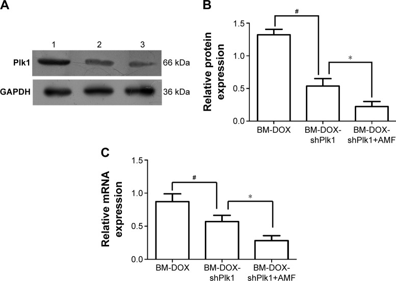
Figure 6.
Plk1 expression driven by the HSP70 promoter was significantly induced by hyperthermia.
Notes: (A) Plk1 protein expression was detected by Western blot. Lane 1, BM-DOX group; lane 2, BM-DOX-shPlk1 group; lane 3, BM-DOX-shPlk1+AMF group. (B) Relative band intensity of Plk1 of BM-DOX, BM-DOX-shPlk1, and BM-DOX-shPlk1+AMF groups compared to the intensity of GAPDH protein in each group. Each value is represented as mean ± SD (n=6). #P<0.05 compared with BM-DOX group. *P<0.05 compared with BM-DOX-shPlk1 group. (C) Relative amounts of Plk1 mRNA expression of BM-DOX, BM-DOX-shPlk1, and BM-DOX-shPlk1+AMF groups compared to the amounts of β-actin mRNA in each group. Each value is represented as mean ± SD (n=6). #P<0.05 compared with BM-DOX group. *P<0.05 compared with BM-DOX-shPlk1 group.
Abbreviations: AMF, alternating magnetic field; BM, bacterial magnetosome; DOX, doxorubicin; HSP70, heat shock protein 70; GAPDH, glyceraldehyde-3-phosphate dehydrogenase; Plk1, polo-like kinase 1; shPlk1, recombinant eukaryotic plasmid pHSP70-Plk1-shRNA; SD, standard deviation.

