Fig. 2.

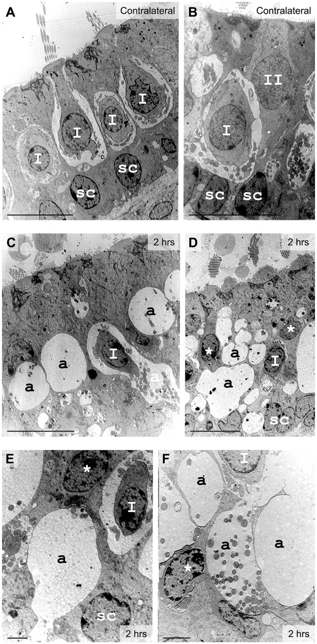
Excitotoxic injury in vestibular utricles observed 2 h after lesion formation with electron microscopy. (A,B) In control tissue, type I hair cells (I) surrounded by calyx nerve endings and cylindrical type II hair cells (II) were identified lying over the nuclear layer of supporting cells (sc). (C-F) In injured inner ear, a few type I hair cells could still be identified from the calyx terminal. Most hair cell types became undetermined (*) because of their distorted morphology induced by large swellings at their base (a). Localization and mitochondrial content was used to characterize the swollen structures as afferent terminals (a). Scale bars: 10 µm (A-D); 2 µm (E,F).
