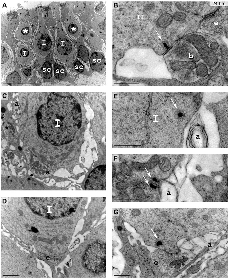
Fig. 5.
Detailed morphology of vestibular epithelia 24 h after the excitotoxic injury. Utricles were observed with electron microscopy. (A) At low magnification, some type I hair cells (I) were identified, whereas other hair cells with an undetermined type remained (*). Sc, supporting cells. (B-G) Nerve terminals were detailed at higher magnification. (B) Typical ribbons (white arrow) facing postsynaptic densities on bouton afferent fibers were observed. II, type II hair cells. (C,D) Bouton-like terminals (a) or efferent terminals (e) unusually connected some type I hair cells. (E,F) Single or multiple ribbons (arrows) faced remaining pieces of calyceal membrane. (G) Typical features of competition between efferent terminals and afferent fibers to contact hair-cell-facing ribbons (arrow) were found. Scale bars: 10 µm (A); 1 µm (C,D); 500 nm (B,E-G).

