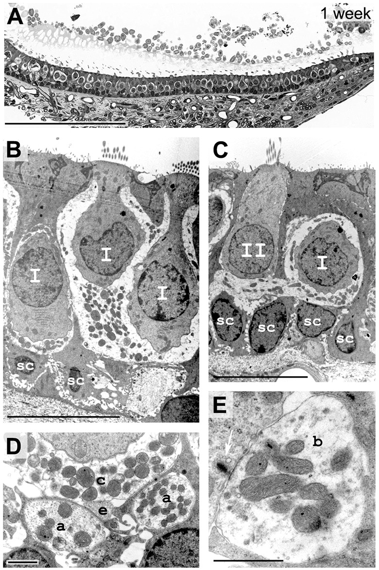
Fig. 6.

Histology of sensory epithelium 1 week after excitotoxic injury. (A) Transverse semi-thin sections of the injured utricle. At 168 h, the lesion shows a normal gross morphology and organization of hair cells and supporting cells, beneath the conjunctive tissue where innervating fibers were observed. (B,C) Electron microscopy shows that morphology of type I (I) and type II (II) hair cells was normal; terminal afferents are present. (D,E) At higher magnification, regular afferent terminals were observed: calyx connect type I hair cells (c), efferent terminals contact calyx afferents (e), afferent fibers are passing (a) and bouton afferent terminals (b) contact facing ribbons (white arrow) of type II hair cells. Scale bars: 100 µm (A); 10 µm (B,C); 1 µm (D,E).
