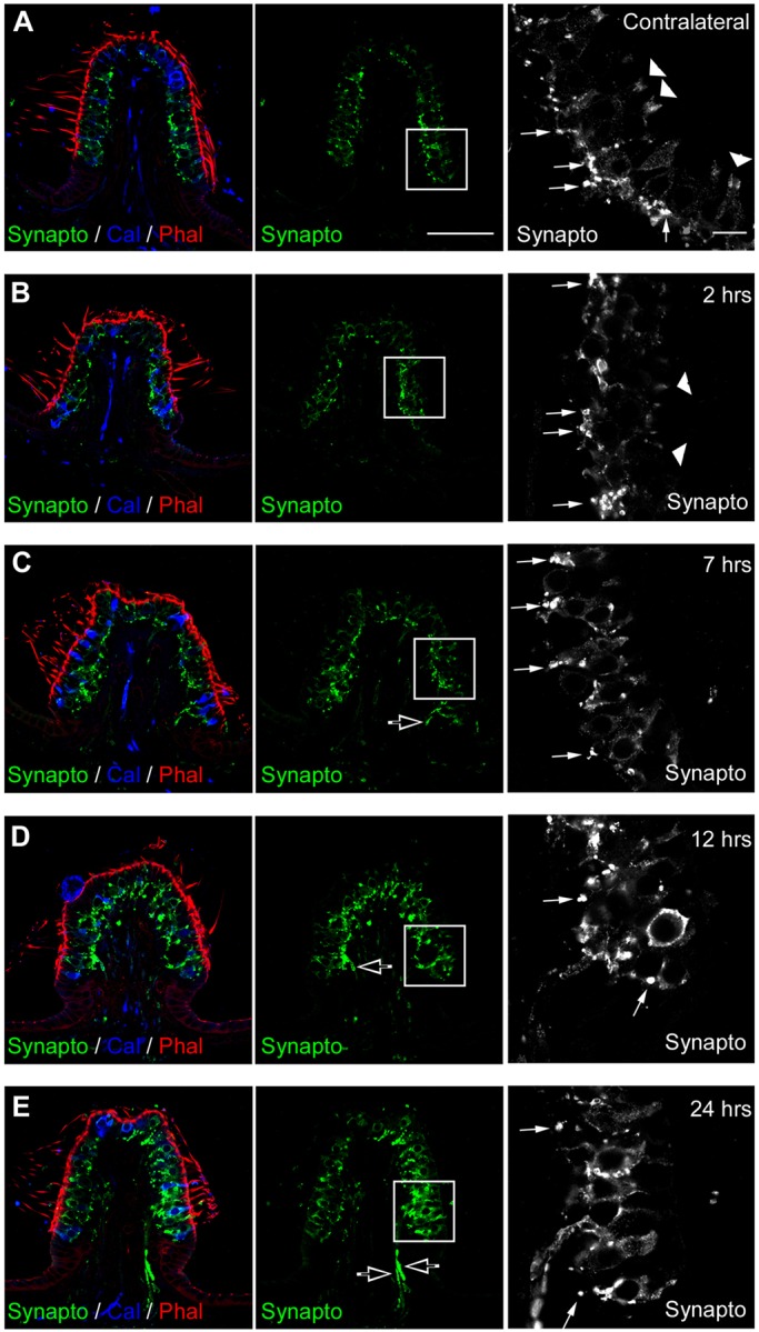
Fig. 7.

Progression of synaptophysin expression over time after excitotoxic injury. Immunolabeling in a transverse section of crista observed by confocal microscopy. (A) In the control ear, counter-labeling of transverse sections with phalloidin (Phal, red) and antibodies against calretinin (Cal, blue) highlight hair bundles of sensory cells, and most type II hair cells and pure calyx of the central zone. Immunolabeling with antibodies against synaptophysin (Synapto, green and white) locates small synaptic vesicles in normal vestibular sensory epithelium. High magnification of the inset (black and white panel) shows precisely that synaptophysin is expressed in efferent terminals (arrows) and at the top of calyces (arrowheads) in this adult tissue. (B-E) Between 2 h and 24 h after the STKK injection, gross morphology of the sensory epithelium was distorted, as evidenced by the actin labeling, and synaptophysin expression changed. (B,C) At 2 h and 7 h, synaptophysin became mostly disorganized, with loss at the top of calyces. (D,E) Between 12 h and 24 h, expression increased in innervating fibers within the conjunctive tissue (open arrows, C-E) and hair cells. Scale bars: 10 µm.
