Fig. 4.

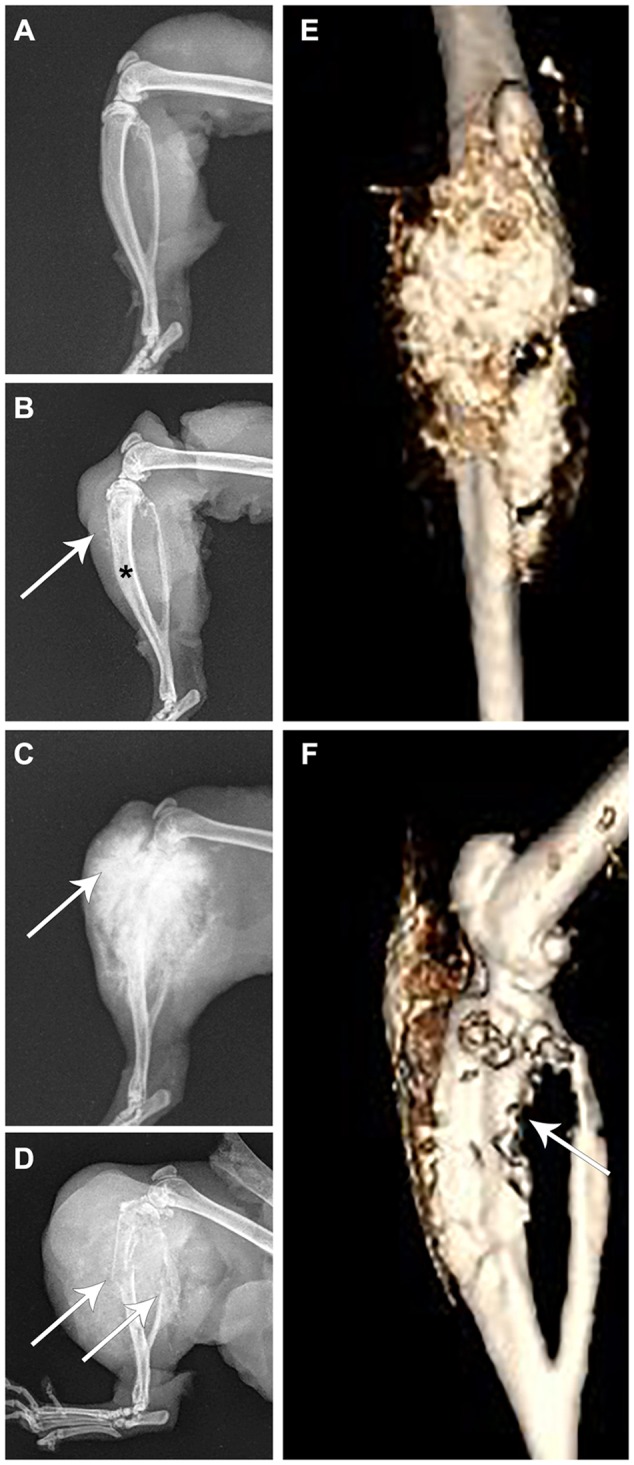
Imaging of osteosarcomas in the F344-Tp53 rat. Rat limbs from four animals. (A) A normal healthy limb. (B,E,F) Different images of the same limb of an animal euthanized for lethargy. (C,D) Limbs from two different rats euthanized because they showed hindlimb swelling and lameness with obvious tumor growth. (A) Unaffected rat tibia and fibula; (B) osteosarcoma of the tibia with loss of normal medullary cavity opacity (asterisk), minimal soft-tissue swelling and early osteoid deposition (arrow); (C) osteosarcoma of the tibia exhibiting a typical ‘sunburst’ pattern of abnormal osteoid deposition in the tumor (arrow); (D) osteosarcoma of the tibia exhibiting pathologic fracture of both the tibia and fibula (arrows), considerable soft-tissue swelling and osteoid deposition; (E) anterior-posterior view in an un-enhanced computed tomography image of the same osteosarcoma shown in B, revealing in more detail the extent of osteoid deposition (gold colored); and (F) lateral view in an un-enhanced computed tomography image of the same limb in B,E exhibiting cortical lysis of the tibia (arrow).
