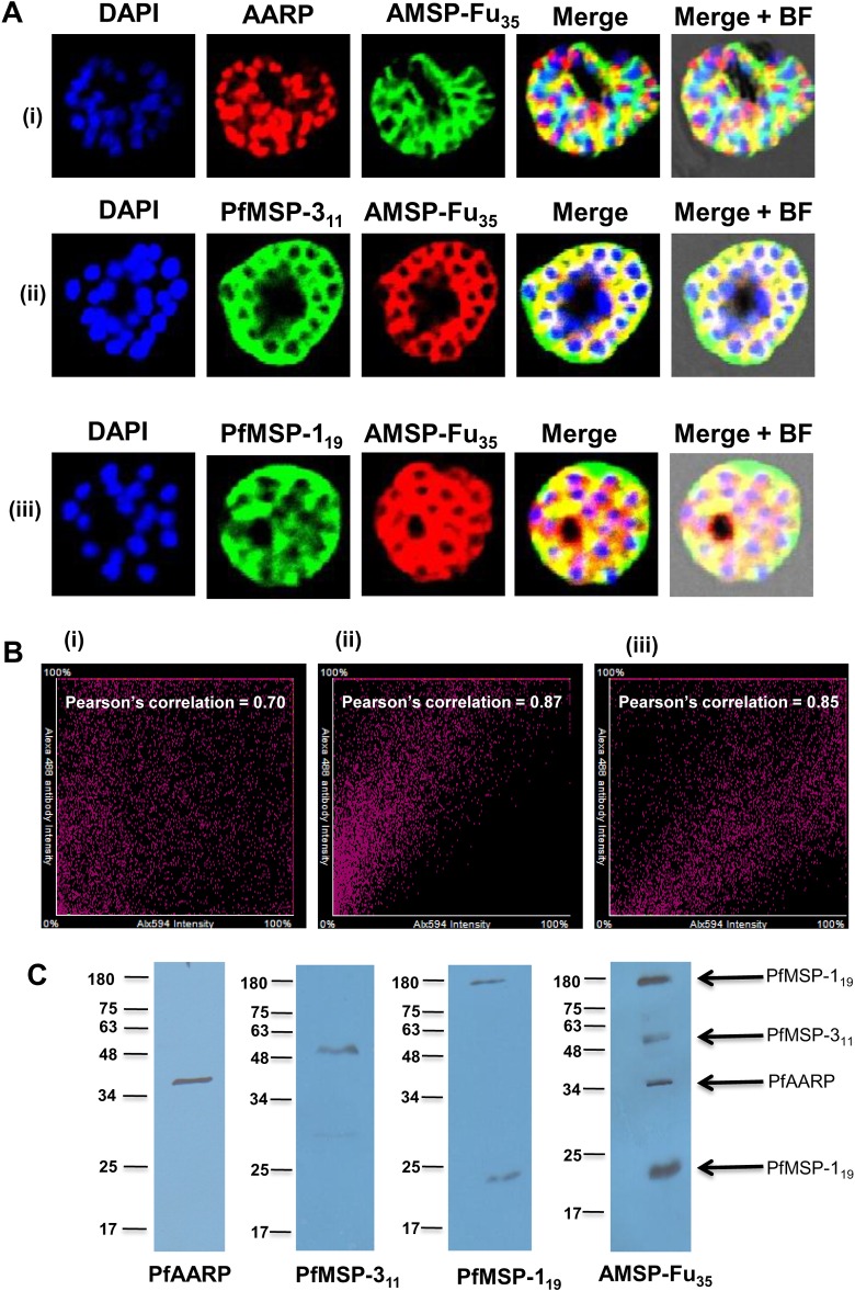
Fig 5. Native conformations of the three individual components in AMSP-Fu35.
(A) IFA was performed with late schizont stage parasites using AMSP-Fu35 sera and co-localization was checked by co-immunostaining with sera against individual components (anti-PfMSP-119, PfAARP and PfMSP-311). The corresponding images stained with DAPI, which stains nuclear DNA; merged fluorescence images and bright field images are also shown. (B) Pearson’s correlation graphs for the co-immunostained slides, (i) AMSP-Fu35 and PfAARP, (ii) AMSP-Fu35 and PfMSP-311 and (iii) AMSP-Fu35 and PfMSP-119, are shown and high correlation values showed co-localization. (C) Antibodies to AMSP-Fu35 react with proteins corresponding to PfAARP, PfMSP-119 and PfMSP-311 in immunoblots of schizont stage parasite lysate from 3D7. Parallel immunoblots of the same lysate probed with anti-AMSP-Fu35, PfAARP, PfMSP-311 and PfMSP-119 sera confirmed the proteins detected by the fusion sera.

