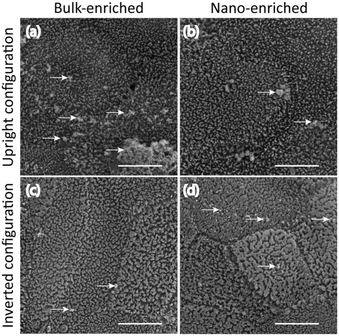
Fig 5. SEM micrographs of human intestine in vitro cell models exposed to gum-E171 nano- or submicron-enriched fractions.

(a) Epithelia exposed to 1 μg/mL of submicron-enriched fraction in the upright configuration resulted in a large number of particles (white arrows) decorating the surface of the epithelium after 7 minutes of exposure. (b) However, exposing replicate samples to the nano-enriched fraction with 1 μg/mL for 7 minutes in the upright configuration resulted in few particles adhered to the epithelial surface. (c) Inverting the epithelium and subsequently exposing the cells with 1 μg/mL of the submicron-enriched fraction for 7 minutes resulted in few particles adhered to the epithelial surface. (d) However, exposing replicate samples in the inverted configuration to 1 μg/mL of the nano-enriched fraction for 7 minutes resulted in relatively more particles adhered to the epithelial surface. All images are shown at identical magnification. Scale bar is 5 μm.
