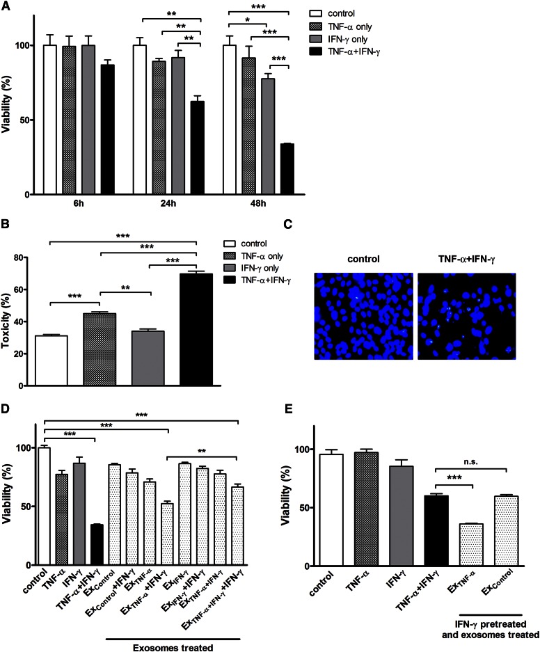
Fig. 1.
Cytotoxic effects of Th1 cytokines (i.e., TNF-α and IFN-γ) and exosomes derived from cytokine-treated cultures on HOG cells. HOG cells were treated with TNF-α (100 ng/ml), IFN-γ (100 ng/ml), or both in combination under serum-free conditions. A: Cell viability of the cytokine-treated cultures after 6, 24, and 48 h was determined by MTT assay, as described in the Materials and Methods. The results expressed as average values are presented as percent of control (untreated cultures) set at 100% ± SEM; *P < 0.05, **P < 0.01, ***P < 0.001; N = 3. B: Cytotoxic effect of the cytokines on HOG cells treated as above was quantified by LDH assay. The results expressed as average values are presented as percent of LDH released from control cultures lysed with Triton X-100 ± SEM; **P < 0.01, ***P < 0.001; N = 3. C: Morphological evidence for apoptotic cells, i.e., Hoechst-stained fragmented DNA in cultures exposed to cytokine combination. D: Exosomes prepared from the media derived from untreated cultures (Excontrol) as well as those treated with TNF-α, IFN-γ, and their combination (i.e., ExTNF-α, ExIFN-γ, and ExTNF-α+IFN-γ, respectively) were used for testing their effects on fresh cultures of HOG cells in the presence and absence of simultaneously added IFN-γ. Cell viability was determined by MTT assay after 48 h and the results are expressed as above; ***P < 0.001; N = 3. E: Another fresh set of HOG cultures were pretreated with IFN-γ (100 ng/ml) for 6 h, washed, and then exposed to either ExTNF-α or Excontrol for 24 h. Cell viability was determined by MTT assay and the results expressed as above; ***P < 0.001; n.s., not significant; N = 3.

