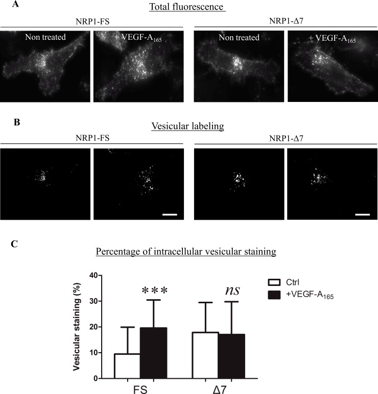
Fig 8. NRP1-FS and NRP1-Δ7 are differentially relocalized after VEGF-A165 stimulation.
PC3 clones induced (dox at 200 ng/ml) to express FLAG-tagged NRP1-FS or NRP1-Δ7 were serum-starved for 24h before exposure or not to VEGF-A165 for 10 minutes. Immunofluorescence analyses were performed using a FLAG antibody and imaging parameters were set to visualize (A) the total amount of tagged-NRP1 or (B) only NRP1 associated with intracellular vesicles in the same cells as in (A). Quantification of the intracellular vesicular NRP1 was performed as described in Materials and Methods. Results are expressed as the mean ± SD (n = 69, FS, 0 VEGF; n = 110, FS +VEGF; n = 77 Δ7, 0 VEGF and n = 72, Δ7, +VEGF; ***: p<0.001, Mann-Whitney U test)—Scale bar = 10 μm.

