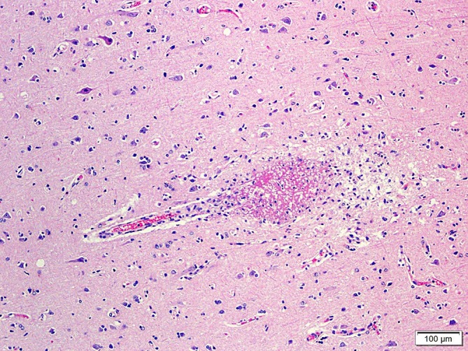
Figure 4. Photomicrography of the brain showing capillary vessel destroyed by micro thrombus. The pineal gland showed numerous full-formed cysts filled with amorphous, acellular, eosinophilic material of unknown origin, which is consistent with the diagnosis of simple pineal cysts. The central nervous system (CNS) microemboli, in contrast with those in the renal glomeruli, were of fibrinous origin and showed no bacteria or inflammatory cells.

