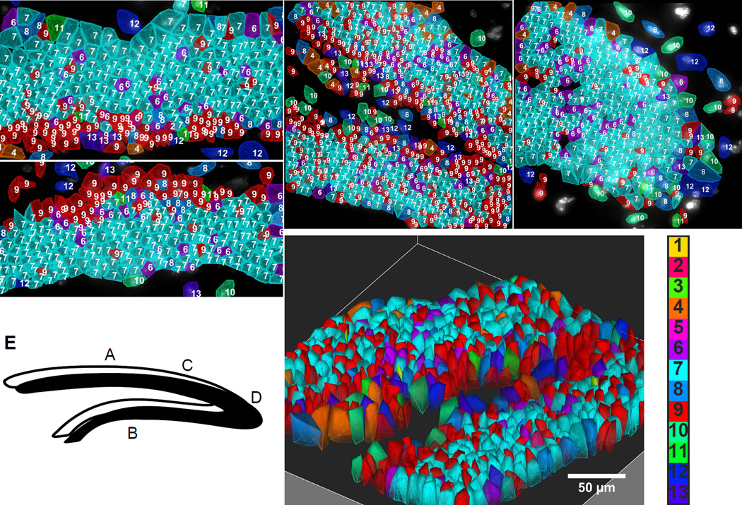
Fig. 4. Spatial layering of cell classes in the Dentate Gyrus (DG).
A–B. Suprapyramidal and infrapyramidal blades of DG. Cells of the subgranular zone and granule cells are arranged in lamina layers in mirror symmetric patterns on the upper and lower blades. C. The SGZ stays on the inner layer of the DG fork. D. Cells are patterned in the crest. Numbered color key corresponds to cluster numbers in Fig 3b. E. Letters in the cartoon of DG correspond to images. F. 3D image of the fork region shown in C.

