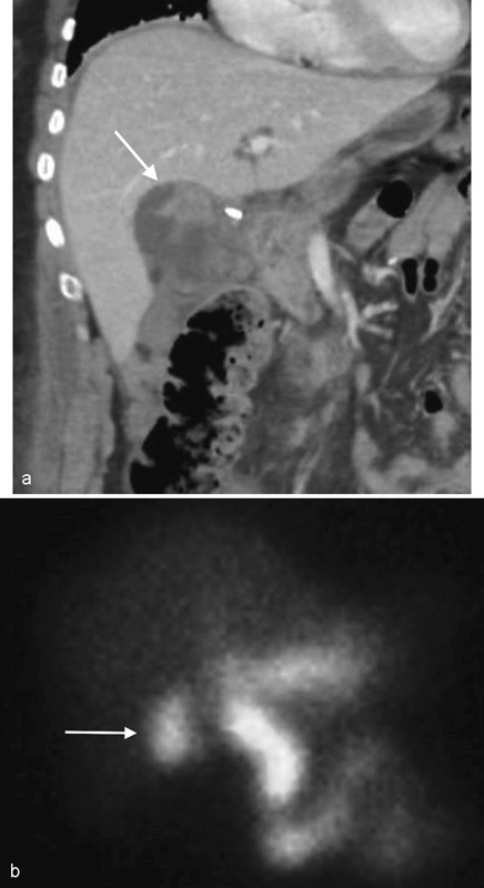
Fig. 1.

A 50-year-old woman with right upper quadrant abdominal pain 2 days after laparoscopic cholecystectomy. (a) Coronal reconstructed image from contrast-enhanced CT abdomen demonstrates a fluid collection of variable density within the gallbladder fossa (arrow). (b) Hepatobiliary cholescintigraphy reveals activity in the region of the cystic duct stump that diffuses into the abdominal right upper quadrant (arrow), consistent with biliary leak.
