
Fig. 2.
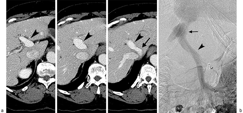
(a) Serial contrast-enhanced axial CT images from caudal (left) to cephalad (right) direction reveal middle hepatic vein (arrow) to left portal vein (arrowhead) shunt. (b) Venous phase image from superior mesenteric arteriogram confirms presence of anatomic portosystemic shunt(arrowhead - portal vein; arrow - hepatic vein).