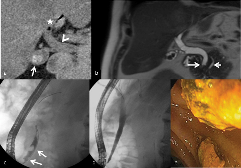
Fig. 1.

A 71-year-old man with abdominal pain and elevated liver function tests. Patient has a remote history of cholelithiasis, sphincterotomy for choledocholithiasis, and refusal of cholecystectomy. (a) Non–contrast-enhanced CT of the abdomen demonstrating cholelithiasis (arrow) with dilated common bile duct (CBD) (arrowhead) and pneumobilia (asterisk). (b) Magnetic resonance cholangiopancreatography showing cholelithiasis and choledocholithiasis (long arrow) with a dilated CBD and prominent pancreatic duct (short arrow) without evidence of pancreatitis. (c) Endoscopic retrograde cholangiopancreatography with multiple filling defects consistent with choledocholithiasis (arrows). (d) Cholangiogram after balloon extraction of stones with resolution of filling defects. (e) Endoscopic image of biliary stones in the duodenum.
