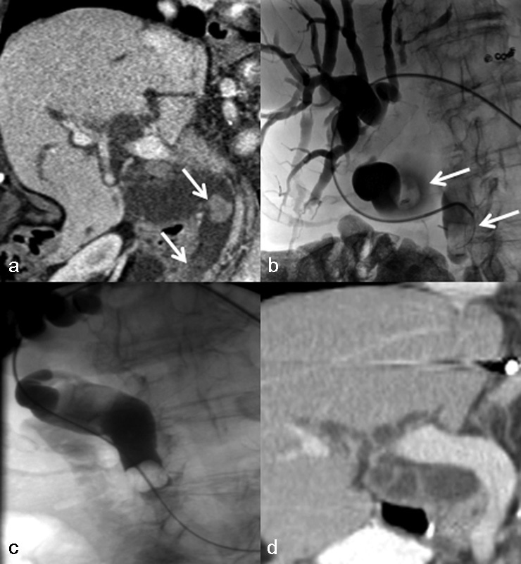
Fig. 2.

A 91-year-old woman with Roux-en-Y anatomy and choledocholithiasis complicated by acute cholangitis. (a) CT scan with oral and intravenous contrast demonstrates stones in the extrahepatic bile duct (arrows), including subtle distal stone, with marked biliary ductal dilation. (b) Percutaneous placement of curved catheter into the extrahepatic bile duct with limited cholangiogram showing dilated intrahepatic and extrahepatic ducts with filling defects corresponding to CT (arrows). (c) After sphincteroplasty, a Fogarty balloon was used to push stones into the small bowel. (d) Follow-up CT scan with resolution of filling defects and decreased biliary ductal dilation.
