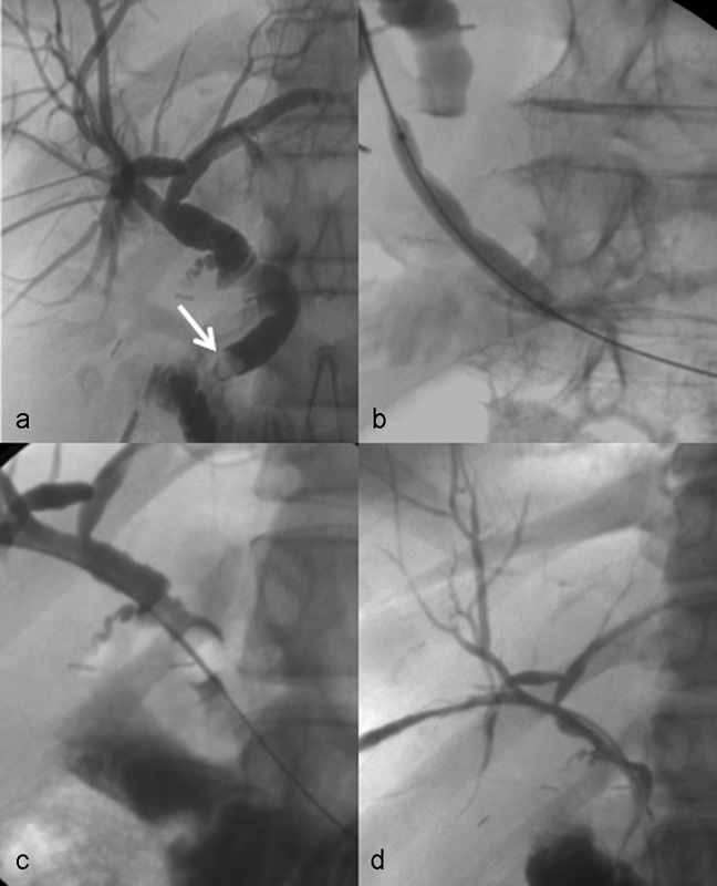
Fig. 3.

A 44-year-old woman with Roux-en-Y anatomy, due to bariatric surgery, with symptomatic choledocholithiasis and failed endoscopic retrograde cholangiopancreatography utilizing double-balloon technique. (a) Percutaneous cholangiogram, with sheath tip in the extrahepatic bile duct, shows postoperative changes of cholecystectomy, with mild biliary ductal dilation and stone in the distal common bile duct (arrow). (b) Balloon sphincteroplasty. (c) Fogarty balloon was used to push stone into duodenum. (d) Delayed sheath cholangiogram showing resolution of biliary ductal dilation and choledocholithiasis with an intact percutaneous tract.
