
FIGURE 1.
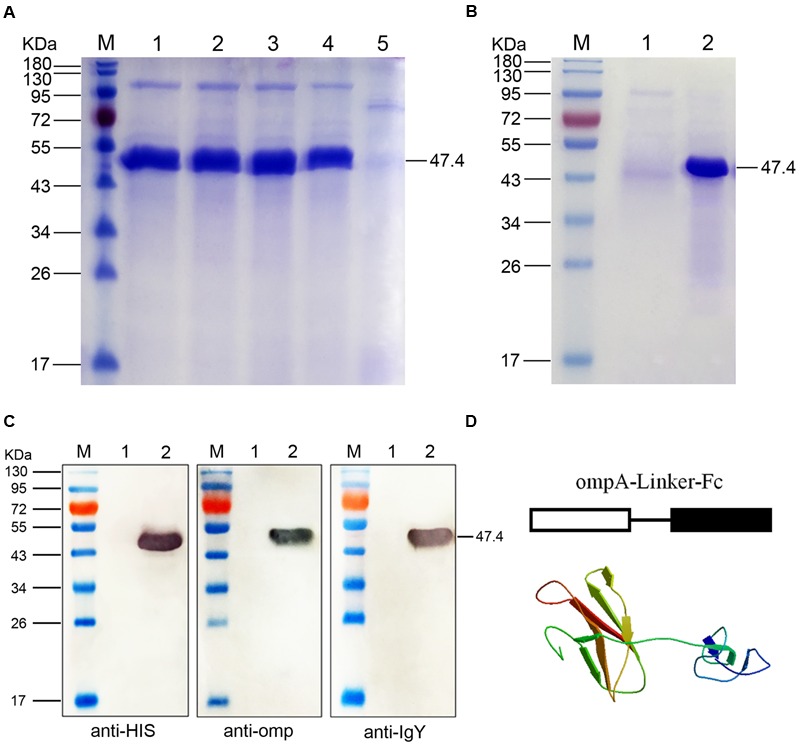
SDS-PAGE and Western blot analyses of the fused ompA–Fc expressed in Pichia pastoris. (A) SDS-PAGE identification of the fusion ompA–Fc at different induction times. M, Page ruler pre-stained protein ladder; lanes 1–4, culture supernatant of P. pastoris transformed with the recombinant pPIC9-ompA–Fc plasmid after 96, 72, 48, and 24 h of methanol induction; lane 5, culture supernatant of P. pastoris transformed with blank pPIC9 vector (negative control). (B) Purification of the fused ompA–Fc. M, Page ruler pre-stained protein ladder; lane 1, purified ompA–Fc; lane 2, culture supernatant after column chromatography. (C) Western blot analyses of the fused ompA–Fc with the anti-His tag antibody, mouse anti-omp polyclonal antibody, and the rabbit anti-chicken IgG (HRP). M, protein molecular size page ruler; lane 1, culture supernatant of P. pastoris transformed with blank pPIC9 vector (negative control); lane 2, culture supernatant of P. pastoris transformed with the recombinant pPIC9-ompA–Fc plasmid at 96 h post induction. (D) Schematic and 3D structure of the fused ompA–Fc.