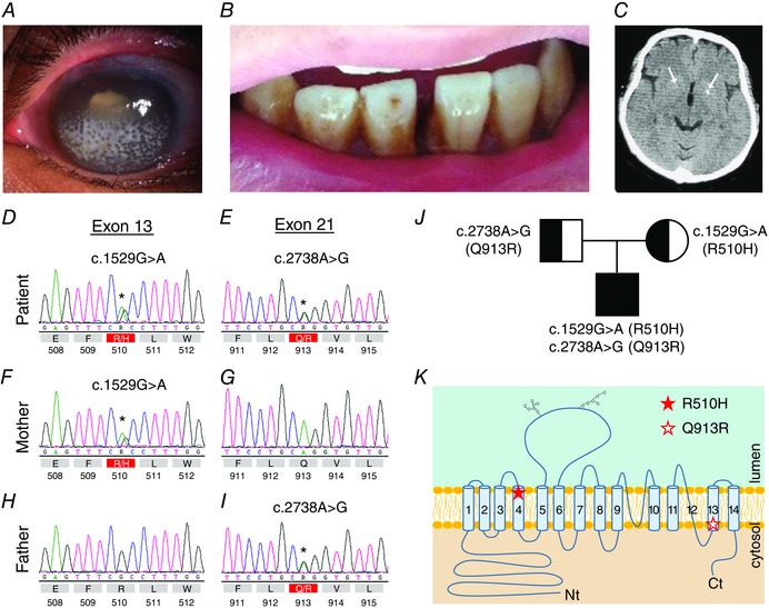
Figure 1. Features of the patient .

A, the patient is blind and exhibits bilateral band keratopathy, cataracts and glaucoma. B, the patient also exhibits signs of enamel hypoplasia and poor teeth alignment. C, computed tomography scan of the patient's head; arrows indicate the location of calcifications. D–I, electropherograms showing the sequence of genomic DNA (prepared from peripheral blood samples) in the vicinity of the mutations for the patient and his parents. The predicted amino acid translation is shown beneath each trace, with mutations indicated in red. J, pedigree of the compound‐heterozygous patient. K, cartoon depicting the predicted topological location of the R510H and Q913R mutations in NBCe1. Note that, in the patient, the mutations are carried on separate SLC4A4 alleles.
