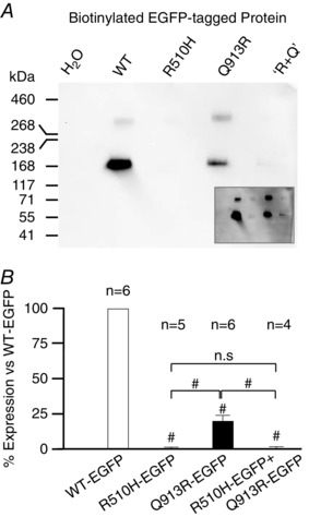
Figure 6. NBCe1‐A‐EGFP protein expression in Xenopus oocytes .

A, representative anti‐EGFP western blot of biotinylated (i.e. extracellular accessible, plasma membrane resident) protein extracts from Xenopus oocytes. ‘R+Q’ denotes cells injected with a 1:1 mixture of R510H‐EGFP + Q913R‐EGFP cRNAs. NBCe1‐A‐EGFP immunoreactivity is barely detectable in lanes 3 and 5 but was evident with longer exposures (A, inset) and exhibits an electrogenic Na/HCO3 cotransport activity that is readily detected by voltage clamp (Fig. 5 C, inset). Protein ladder is HiMark™ pre‐stained protein standard (Thermo Fisher Scientific). B, averaged intensities of immunoreactive bands from blots such as that shown in (A), normalized to the intensity of WT‐EGFP. #Significance vs. WT‐EGFP with 95% confidence using an ANOVA with Tukey's comparison test. #Significance in the same test between the groups indicated. n.s., no significant difference.
