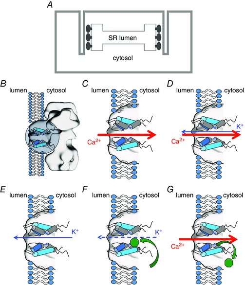
Figure 1. Ionic fluxes and interaction of flecainide in RyR2 .

A, schematic representation of the location and orientation of RyR2 in the cardiac myocyte SR membrane system. The trans‐membrane domain contains the pore‐forming region (PFR) and the large cytosolic domain abuts transverse tubule invaginations of the sarcolemma. B–G, schematic diagrams depicting the open RyR2 PFR (B, indicated by circle) together with relevant cation fluxes and interactions of flecainide (green octagon). Only two of the four PFR monomers are shown for clarity. C, to initiate contraction Ca2+ is released from the SR, flowing down its concentration gradient through the open RyR2 PFR. D, to dissipate the diffusion potential generated by Ca2+ release, a counter current of K+ flows from the cytosol to the SR lumen, at least in part through RyR2. E, the RyR2 blocking parameters used in Yang et al. (2016) are based on data reported in Hilliard et al. (2010) that describe the effects of flecainide on the cytosolic to luminal flux of K+. F, under these conditions flecainide enters the cytosolic cavity of RyR2 and partially occludes the pore. G, the physiologically relevant flux of Ca2+ from the lumen to the cytosol displaces flecainide (Bannister et al. 2015). As a consequence open channel block of RyR2 by flecainide can play no part in the effectiveness of this drug in the treatment of CPVT.
