
Figure 1. Morphology and physiological responses of pancreatic stellate cells (PSCs) .
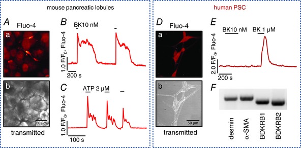
A, mouse pancreatic lobules: a, pancreatic stellate cells (PSCs) stain avidly with the Ca2+‐sensitive dye Fluo‐4; b, transmitted light image of a. B, typical Ca2+ response to 10 nm bradykinin (BK) in mouse PSCs (n = 7). C, typical Ca2+ response to 2 μm ATP (n = 5). D, human pancreatic stellate cells (hPSCs) in culture: a, hPSCs stained with Fluo‐4; b, transmitted light image of a. E, typical responses to bradykinin (BK) in hPSCs (n = 12): 1 μm BK induces a Ca2+ transient, whereas 10 nm does not. F, mRNA expression of desmin, α‐smooth muscle actin, bradykinin receptor B1 and B2 in hPSCs.