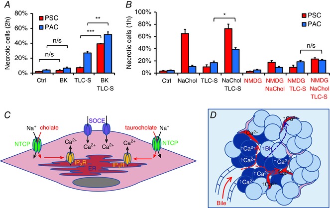
Figure 7. Escalated effects of bile acids in pancreatic lobules .

A, necrosis in mouse PSCs (red) and PACs (blue) treated for 2 h with 10 nm bradykinin (BK), 200 μm taurolithocholic acid 3‐sulfate (TLC‐S) or both (N = 4 for all groups). B, necrosis induced in mouse PSCs (red) and PACs (blue) during 1 h incubation with 5 mm sodium cholate (NaChol), 200 μm taurolithocholic acid 3‐sulfate (TLC‐S) and both bile acids, all in the presence (NaHepes) or absence of extracellular Na+ (NMDG‐Hepes) (N = 3 for all groups). C, proposed mechanism of bile acid‐induced Ca2+ signals in a PSC: bile acids (cholate and taurocholate) enter PSCs via membrane transporters, such as NTCP, and induce IP3R‐mediated Ca2+ release from the ER; depletion of the ER triggers opening of store‐operated Ca2+ entry (SOCE) channels leading to [Ca2+]i overload. D, schematic illustration of Ca2+ events in acute biliary pancreatitis: concentrated bile reaches pancreatic lobules and triggers excessive Ca2+ responses in PACs (blue) and PSCs (red) followed by necrosis; damage to PACs leads to generation of bradykinin, which, in turn, induces Ca2+ responses in PSCs making them more susceptible to bile‐elicited damage.
