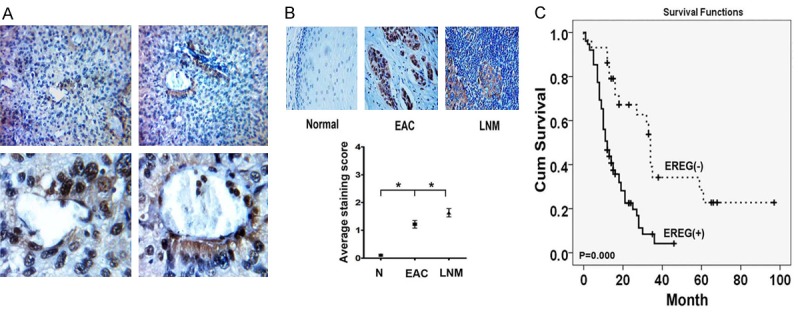
Figure 5.

Expression of EREG in human esophageal carcinoma and Lymph node metastatic tissues. A. Representative images of EREG staining in perivascular regions of esophageal carcinoma tissues. B. IHC analysis of EREG expression in primary and Lymph node metastatic esophageal carcinoma. C. Kaplan Meier survival curve showed correlation between EREG level and overall survival.
