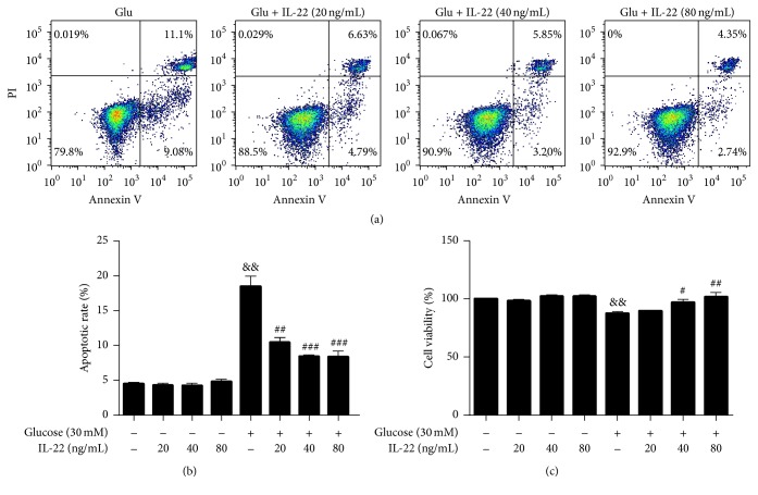
Figure 5.
IL-22 protected HUVECs from glucose-induced injury. Cells were treated with glucose (30 mM) for 12 h, followed by incubation with different concentrations of IL-22 for 24 h. The apoptotic rate was quantified by flow cytometry. Cell viability was analyzed using the CCK-8 assay and the results were compared with those obtained for the control group. Data shown represent mean ± SEM of three independent experiments. && P < 0.01 versus control group; # P < 0.05; ## P < 0.01 versus glucose-treated group; ### P < 0.0001 versus glucose-treated group.

