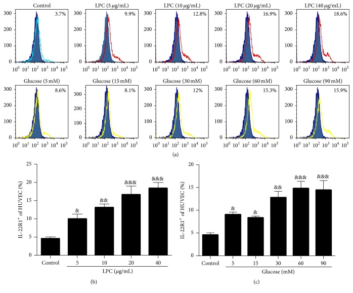
Figure 6.
LPC and high concentrations of glucose increased IL-22R1 expression in HUVECs. Representative flow cytometry histogram of IL-22R1 expression on HUVECs. Surface expression of IL-22R1 (open area) on HUVEC single-cell suspensions in comparison with the isotype control (filled area), as determined by flow cytometry. Green open area represents control group. Red open area represents LPC group. Yellow open area represents glucose group. The percentages of IL-22R1+ cells among the HUVECs were analyzed by using FlowJo. Data shown represent mean ± SEM of three independent experiments. & P < 0.05, && P < 0.01, and &&& P < 0.0001 versus control group.

