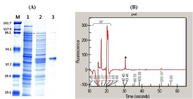
Figure 5.

Protein purification of heterologously expressed NfAXE1 in Escherichia coli. (A) SDS-PAGE with Coomassie Blue staining of proteins at various stages of purification. M, Molecular markers (kDa). Lanes: 1, Cells harboring pET28a as control; 2, crude cells harboring pET28a-NfAXE1 after IPTG induction; 3, purified recombinant NfAXE1. (B) SP, Internal standard signal peaks; *, purified target protein peak analyzed using the microfluidic system. NfAXE1, Neocallimastix frontalis PMA02 acetyl xylan esterase; SDS-PAGE, sodium dodecyl sulfate-polyacrylamide gel electrophoresis.
