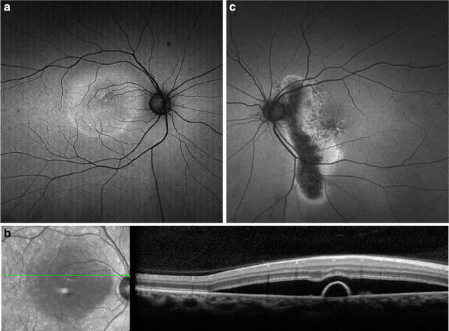
Fig. 8.

Fundus autofluorescence of central serous chorioretinopathy. FAF (a) of a son with acute exacerbation of CSCR shows macular detachment due to acute CSCR, with hyper-autofluorescent material at the margin and inferior region of the detachment. SD-OCT (b) of the lesion shows a serous retinal detachment associated with a small pigment epithelial detachment. FAF of the patient’s asymptomatic father (c) incidentally revealed an atrophic hypo-autofluorescent gravitational tract from chronic inactive CSCR with hyper-autofluorescent margins
