Figure 1.

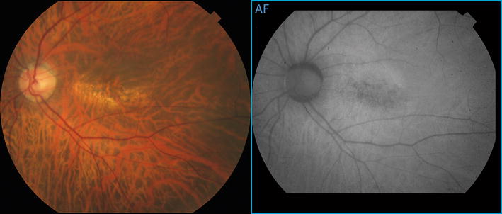
Color photograph and fundus autofluorescence images of the posterior pole of the left eye show a rectangular area of mottled retinal pigment epithelial atrophy arranged with its long axis aligned horizontally.

Color photograph and fundus autofluorescence images of the posterior pole of the left eye show a rectangular area of mottled retinal pigment epithelial atrophy arranged with its long axis aligned horizontally.