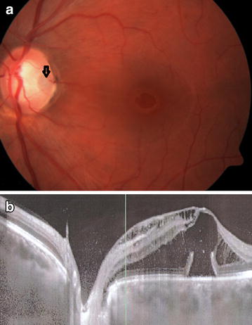
Fig. 1.

a Fundus photograph of the left eye of a 31 year old man with ODP-M. A temporal ODP is noted (arrow). VA was 20/200. b A horizontal OCT scan through the optic disc and fovea, showing an abundance of intraretinal and subretinal fluid, extending towards the ODP. ODP optic disc pit, ODP-M optic disc pit maculopathy, VA visual acuity and OCT optical coherence tomography.
