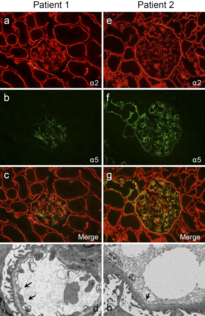
Figure 2.
Renal biopsy findings in X-linked Alport syndrome with a novel mutation. Patient 1 shows (a) a normal pattern of the alpha 2 chain of type IV collagen in the GBM and (b) segmental and mosaic patterns of the alpha 5 chain of type IV collagen in the GBM and the absence of staining in Bowman’s capsule and the TBM. (c) The merged findings of the alpha 2 and 5 chains of type IV collagen are shown. (d) Electron microscopy reveals a split lamina densa (arrows) and thin GBM. Patient 2 shows (e) a normal pattern of the alpha 2 chain of type IV collagen in the GBM and (f) a mosaic pattern of the alpha 5 chain of type IV collagen in the GBM and TBM. (g) The merged findings of the alpha 2 and 5 chains of type IV collagen are shown. (h) Electron microscopy reveals a split lamina densa (arrow). GBM: glomerular basement membrane, TBM: tubular basement membrane

