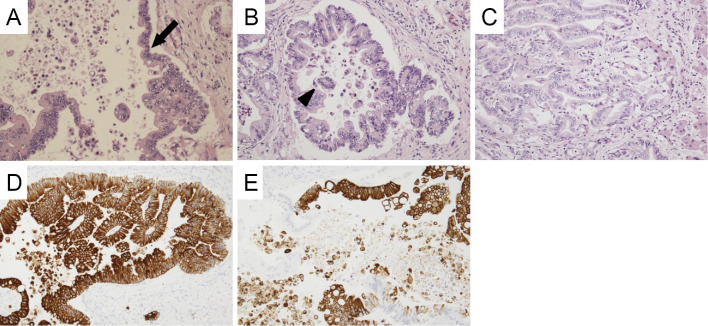
Figure 3.
Microscopic findings of the hepatic tumor. Adenocarcinoma is seen spreading along the bile duct wall (arrow) (A). The tumor shows stromal invasion forming irregular tubular structures with or without papillary projection (B, C). Exfoliation of carcinoma cell clusters (arrowhead) is observed in the ductal space (B). Carcinoma cells are diffusely positive for CK7 (D) and partially positive for CK20 (E).

