Figure 4.
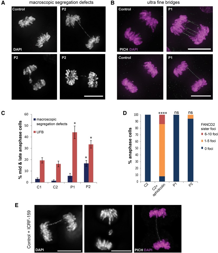
Condensin patient mutations lead to decatenation failure at mitosis. (A–C) Chromosome segregation is impaired in patient P1 (NCAPD2) and P2 (NCAPD3) primary fibroblasts. Representative images of chromatin bridges and lagging chromosomes/chromosome fragments were detected with DAPI stain (macroscopic chromosome segregation defects) (A), and ultrafine DNA bridges (UFBs) were detected by the presence of PLK1-interacting checkpoint helicase (PICH) and absence of DAPI stain (B). Bars, 10 µm. (C) Quantification of macroscopic chromosome segregation defects (defined as DAPI-positive chromatin bridges, lagging chromosomes, or chromosome fragments) and UFBs, scored in anaphase C1 and C2 and patient P1 and P2 fibroblasts. Increased numbers of mid and late anaphase cells with UFBs are seen in NCAPD2 patient P1 and NCAPD3 patient P2 cell fibroblasts (experiments ≥ 3; n > 50 anaphases per sample per experiment), and patient P2 fibroblasts also had elevated macroscopic chromosome segregation defects compared with C1 and C2 fibroblasts (experiments ≥ 3, n > 100). Error bars indicate SEM. Two-tailed t-test, (*) P ≤ 0.05 versus C1. (D) UFBs in condensin patient cells do not arise from late replicating intermediates. Quantification of the number of FANCD2 sister foci during anaphase in C2 and patient P1 and P2 fibroblasts. C2 fibroblasts treated with 0.1 µM aphidicolin for 16 h to induce late replicating intermediates through replication stress were also included as a positive control. n = 50 cells; experiment = 1. (E) Chromosome decatenation failure due to topoisomerase II α inhibition in control fibroblasts results in chromosome segregation defects identical to those observed in condensin patient cells. Representative images of bulky chromatin bridges and lagging chromatin detected with DAPI stain and UFBs detected by PICH stain. Bars, 10 µm. C2 fibroblasts were treated with 5 µM ICRF-159 for 24 h.

