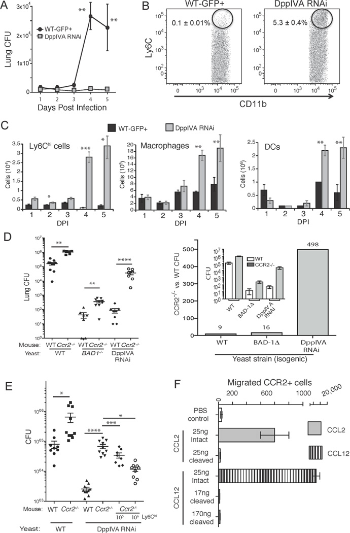
Fig. 2. The inflammatory response to yeast that express DppIVA and the role of CCR2+ monocytes.
(A) Lung CFU of mice infected with isogenic DppIVA-sufficient and –deficient yeast over a 5-days. (B) Percentage of Ly6Chi CCR2+ cells in the lungs 3 days post-infection (events shown are those from the CD11b+ Ly6G− gate) and (C) total number of Ly6Chi cells in lungs. Also shown are the numbers of lung exudative Mϕs and dendritic cells (DC). Data are mean±SEM of 5 mice/group and representative of 3 experiments. (D) Wild-type and Ccr2−/− mice were infected with isogenic yeast and analyzed for lung CFU 2 weeks post infection (left). The mean CFU data (inset) is expressed as fold-change between Ccr2−/− and wild-type mice (right). (E) Ly6Chi cells recruited to lungs and regional lymph nodes of wild-type mice (infected with DppIVA RNAi yeast) were transferred into Ccr2−/− mice infected with DppIVA-silenced yeast. Lung CFU is shown 7 days post infection. Data are mean±SEM of 8–10 mice/group. Results are representative of 2 experiments. (F) Transwell migration of CCR2+ cells in response to intact or fungal DppIVA-cleaved C-C chemokines. Migration was quantified after 16–24 hr of incubation. Results are representative of two experiments. *p<0.05, **p<0.01, ***p<0.001, ****p<0.0001. (See also Figure S1)

