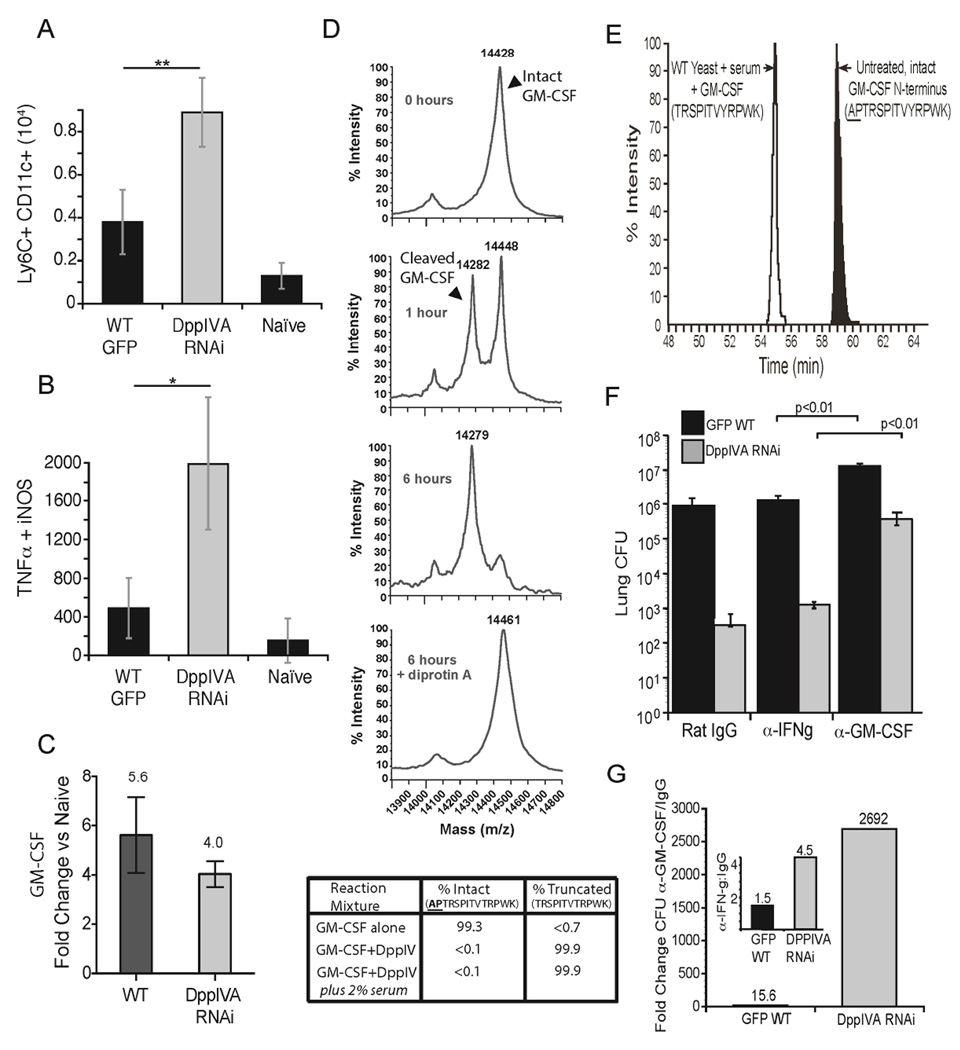
Fig. 3. Action of yeast DppIVA on GM-CSF and impact on evolution of infection.
Number of monocyte-derived DC (CD11c+, Ly6Chi cells) (A) and TipDC (TNF-α, NOS2+, CD11c+, Ly6Chi) (B) in the lungs 4 days post-infection. *p<0.05, **p<0.01. (C) Fold increase in transcript of GM-CSF measured by RT-PCR in lung homogenate 2 days post infection compared to naïve mice. Data in panels A–C are mean±SEM of 3–5 mice/group and are representative of 2 experiments. (D) MS analysis of GM-CSF cleavage by fungal rDppIVA at 0, 1, and 6 hr of incubation alone or with DiprotinA. Table shows extent of removal of N-terminal two amino acids (AP) by rDppIVA in buffer alone or with 2% serum. (E) Wild-type yeast were cultured in vitro in medium with GM-CSF and 2% mouse serum. After 72 hr, GM- CSF was collected by chromatography and analyzed by MS for residues at the N-terminus (open peak) vs. untreated GM-CSF (shaded peak). (F) Mice received rat IgG or neutralizing α-GM-CSF or α-IFN-γ and were infected with yeast strains, and lung CFU enumerated as above in panel E. (G) Fold change in CFU between GM-CSF neutralized vs. rat IgG treated mice is shown for each yeast strain. Inset shows fold-change for rat IgG-treated vs. IFN-γ neutralized mice. Numbers above bars are fold-change. Data in panels E+F are mean ± SEM of 8–10 mice/group, and are representative of 2 experiments. (See also Figure S4).

