Fig. 2.

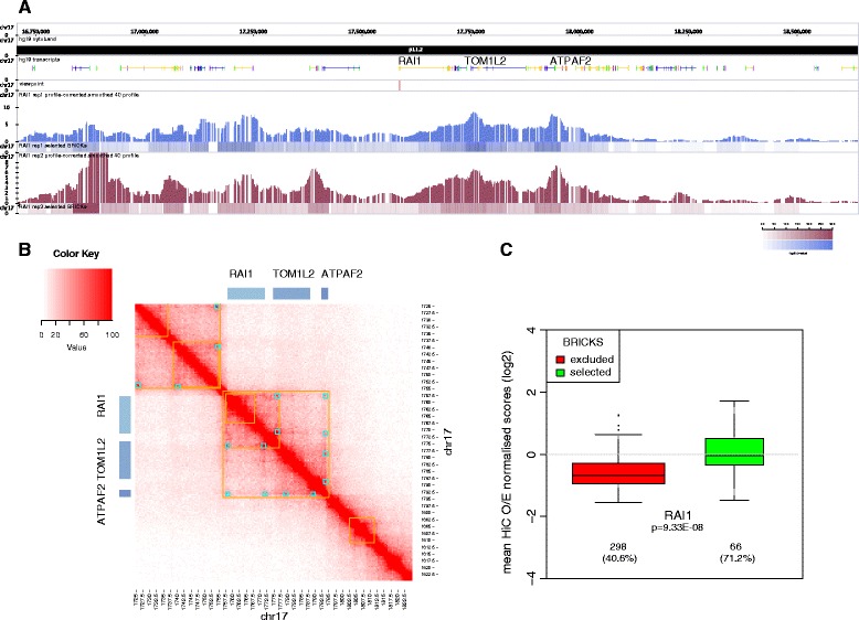
4C interactions profile of RAI1 and comparison with Hi-C interactions profiles locally and globally. a (Panels from top to bottom). Transcripts: The structure of the transcripts mapping within human 17p11.2 cytoband from approximately 16.5 Mb to 18.5 Mb are indicated, in particular those of the RAI1, TOM1L2, and ATPAF2 genes. Viewpoint: The red tick shows the mapping position of the RAI1 viewpoint used in the 4C experiments. PC/BRICKs: Smoothed and profile-corrected 4C signal (upper part of each panel) and BRICKs (lower part) identified for each replicate (blue and burgundy). The corresponding BRICKs significance heatmap color legend is shown in the bottom right corner. b High resolution Hi-C chromosome conformation capture results obtained with the GM12878 LCL within the chromosome 17 17.25-18.22 Mb window (5 kb resolution). Yellow and light blue squares highlight the contact domains and peaks identified in [47], respectively. The position of the RAI1, TOM1L2, and ATPAF2 genes is indicated. c Distribution of Hi-C scores in selected (FDR1%) versus non-selected BRICKS (FDR10%). Virtual 4C-seq tracks were generated for the RAI1 viewpoint from the GM12878 Hi-C results published in [47] (5 kb resolution) by extracting the Hi-C vectors from the KR normalized observed/expected matrices. BRICKS found with the viewpoint were quantified by the mean Hi-C signals. The p value of the two-sided t-test is reported for the comparison, together with the number of Hi-C bins and the % of non-NA bins
