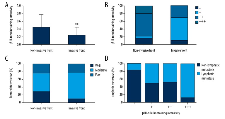
Figure 2.
Positive staining (A) and staining intensity of βIII-tubulin (B) and tumor differentiation (C) were significantly different in patients in the invasive and non-invasive front groups. Staining intensity of βIII-tubulin was significantly associated with lymphatic metastasis in the non-invasive front group (D). ** p<0.01 vs. the non-invasive front group.

