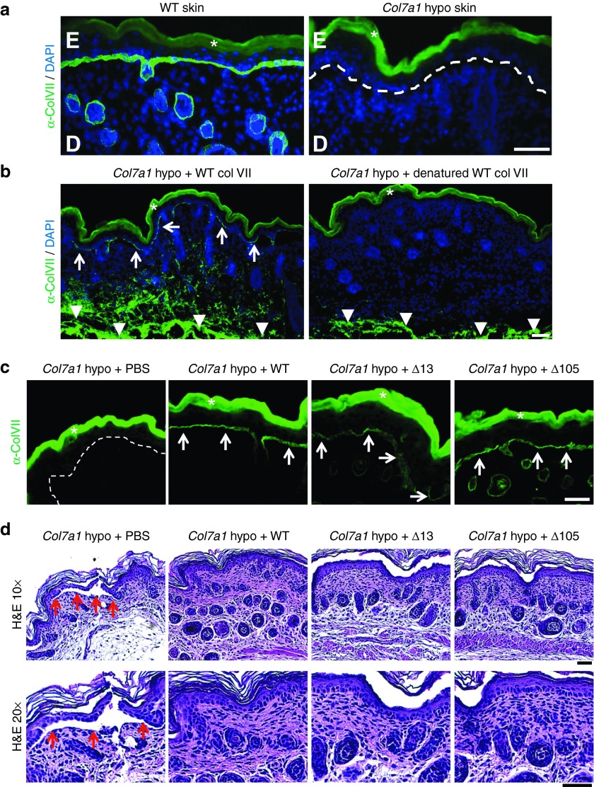
Figure 6.
Collagen VII variants can be deposited at the dermal-epidermal junction in RDEB mice. (a) Skin tissue sections from WT and Col7a1 hypomorphic mice immunostained for collagen VII (green). While WT skin exhibits a strong signal for collagen VII at the dermal-epidermal junction zone, minimal collagen VII deposition is seen in Col7a1 hypomorphic mice (white dotted line). (b) Skin sections stained for human collagen VII (green) after intradermal injection of Col7a1 hypomorphic mice with 25 µg of WT collagen VII and heat denatured WT collagen VII. The staining reveals that only correctly folded collagen VII is able to translocate to the dermal-epidermal junction after injection (white arrows). Note that the injected collagen VII is clearly visible in deeper dermal areas in both skin sections (white arrowheads). (c) Skin sections one day after intradermal injection of Col7a1 hypomorphic mice with 10 µg of WT, Δ13, and Δ105 collagen VII stained for human collagen VII (green). The staining reveals that deletion of amino acids encoded by exon 13 or 105 does not affect the ability of collagen VII to translocate to the dermal-epidermal junction zone compared to WT collagen VII (white arrows). (d) H&E staining of skin sections as in (c). The histological analysis reveals that collagen VII variants lacking amino acids encoded by exon 13 or 105 promote dermal-epidermal stability equally well as WT collagen VII. While vehicle injected mice show areas of dermal-epidermal separation (red arrows), such separations are not visible in mice which had received 10 µg of WT, Δ13, or Δ105 collagen VII. Bar = 50 µm, D = dermis, E = epidermis, in a and b nuclei visualized by 4′,6-diamidino-2-phenylindole (blue). White asterisks indicate autofluorescence of the epidermis.

