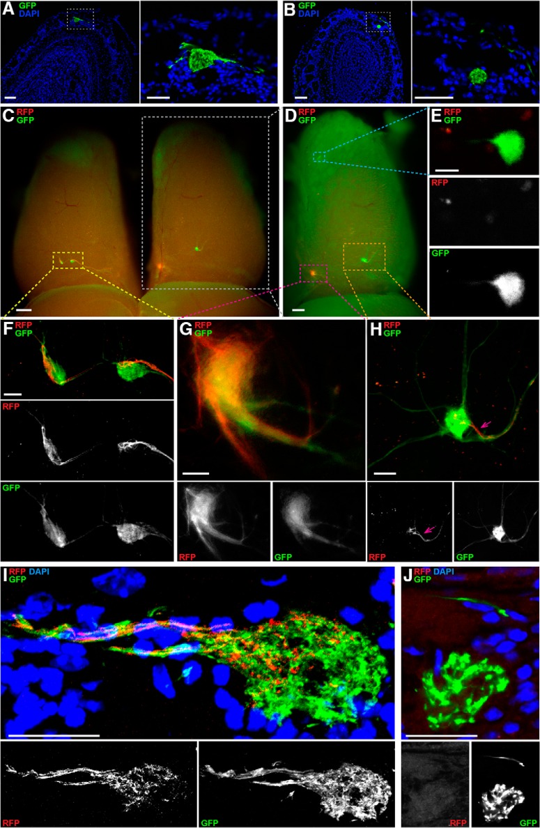
Figure 4.
Confocal and whole-mount imaging of M71 glomeruli in triple-mutant NMCZ and quadruple-mutant NMRCZ mice. A, B, Coronal 12-µm sections of bulbs of NMCZ mice at PD21 imaged with a Zeiss LSM 710 confocal microscope, using the intrinsic fluorescence of GFP and counterstaining of nuclei with DAPI. The box indicated with a white stippled line in the left images is magnified in the right images. A, Ectopic dorsal glomerulus, large. B, Ectopic anterior glomerulus, small and located deeper in the glomerular layer. C–H, Whole-mount bulbs of an NMRCZ mouse at PD59 imaged in wide-field on a Nikon SMZ25 stereofluorescence microscope. C, Dorsal view of the left and right bulbs, with the left bulb exhibiting configuration VI, and the right bulb, configuration V. Not all glomeruli are visible in this view. D, The right bulb from C (white box) is tilted to the right to provide a better view of the medial aspect and demonstrate the configuration consisting of an ectopic anterior glomerulus (blue box), a medial glomerulus (red box), and an ectopic dorsal glomerulus (orange box). E, Magnified view of the ectopic anterior glomerulus and consisting of GFP+ axons, as shown in the blue box in D. F, Magnified view of the two dorsal glomeruli of the left bulb, in the yellow box in C. The left glomerulus exhibits a mixed, comparable contribution of GFP+ and RFP+ axons. The right glomerulus consists mostly of GFP+ axons, with a compartment of RFP+ axons. G, Magnified view of the mixed GFP+ RFP+ medial glomerulus of the right bulb, as shown in the red box in D. H, Magnified view of the ectopic dorsal glomerulus consisting predominantly of GFP+ axons, as shown in the orange box in D. A small compartment of this glomerulus is innervated by an axon bundle containing also RFP+ axons (pink arrow). I, J, 12-µm sections of bulbs of NMRCZ mice at PD56 and PD70, respectively, imaged with a Zeiss LSM 710 confocal microscope, using the intrinsic fluorescence of GFP and RFP and counterstaining of nuclei with DAPI. I, Ectopic dorsal glomerulus consisting of RFP+ and GFP+ axons. J, Magnified view of an ectopic-anterior glomerulus consisting of GFP+ axons. Scale bars, A left, 100 µm; A right, 50 µm; B left, 100 µm; B right, 50 µm; C and D, 300 µm; E, 20 µm; F, 50 µm; G, 25 µm; H, 50 µm; and I and J, 25 µm.

