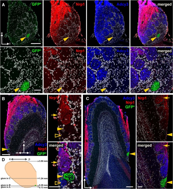
Figure 6.
Posterior glomeruli in the vicinity of Gucy1b2+ glomeruli can be Nrp1+ or Nrp1–. Fluorescence images of three horizontal sections of a bulb of a homozygous Gucy1b2-IRES-tauGFP mouse at 4 weeks were taken with a Zeiss LSM 710 confocal microscope. Each of the three Gucy1b2+ glomeruli is indicated with a yellow arrowhead. A, Section at a very ventral level, 0.13 mm from the bottom of the bulb, as shown schematically in D. Intrinsic GFP fluorescence (GFP*) is combined with immunofluorescence for Nrp1 (red) and Adcy3 (blue). Nuclear staining with DAPI is in white. Merged is all colors together. The top four panels show the entire section. The bottom four panels show high-magnification views of an area surrounding the GFP+ glomerulus. This glomerulus is Adcy3– and Nrp1–. The posterior part of the bulb at this very ventral level is devoid of Nrp1 immunofluorescence but contains numerous glomeruli that are Adcy3+. B, Section at a more dorsal level, 0.29 mm from the bottom of the bulb. The image on the left shows the entire section. The two panels on the right show high-magnification views of an area surrounding the GFP+ glomerulus. This extremely posterior GFP+ glomerulus is Adcy3– and Nrp1–. The glomerulus anterior to the GFP+ glomerulus is Adcy3+ and Nrp1+ (yellow arrow), but the glomerulus posterior to the GFP+ glomerulus is Adcy3+ and Nrp1– (unfilled yellow arrowhead). C, Section at an intermediate dorsal-ventral level, 1.24 mm from the bottom of the bulb. The image on the left shows the entire section. The two panels on the right show high-magnification views of an area surrounding the GFP+ glomerulus. This very posterior GFP+ glomerulus is Adcy3– and Nrp1–. The glomeruli anterior to it are strongly Adcy3+ and Nrp1–, and the most anterior glomerulus (indicated with an arrow) is Adcy3+ and weakly Nrp1+. D, Schematic diagram of a medial view on the bulb. The positions of the three glomeruli shown in A–C are indicated. Scale bars, A, top 200 μm, bottom 50 μm; B and C, left 200 μm, right 50 μm. a, anterior; p, posterior; m, medial; l, lateral; glom, glomerulus.

