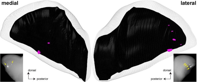
Figure 9.
Example of a three-dimensional reconstruction of a bulb of a PD14 transgenic mouse expressing rat OR I7 from a mouse MOR23 promoter and gap-YFP. The right bulb of a mouse was reconstructed in 3D after epifluorescence whole-mount imaging with a Nikon SMZ25 stereomicroscope. The images of the medial and lateral aspects of this bulb are the same as in Figure 8, right bottom, and reproduced here to facilitate comparison with the 3D reconstruction. There are two labeled glomeruli in the medial aspect (compared to two in the epifluorescence whole-mount image, yellow arrows), and five labeled glomeruli in the lateral aspect (compared to four in the epifluorescence whole-mount image, yellow arrows).

