
Figure 4. TSCs and Blastocysts Endocytose Primary Uterine Epithelial Cells In Vitro.
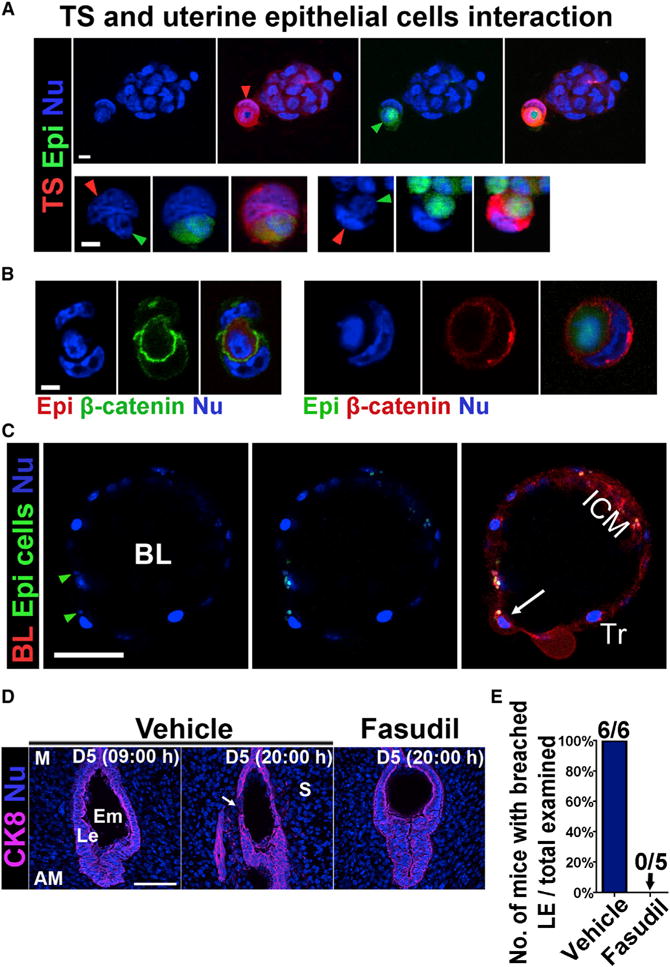
(A) TSCs engulf primary epithelial cells. Red arrowhead points toward the nucleus of one TSC (red), whose nucleus is squeezed to a crescent shape by an epithelial cell (green). The green arrowhead points toward the engulfed epithelial cell. The bottom panels show two other engulfed epithelial cells (green) by TSCs (red). Scale bar represents 10 μm.
(B) Immunofluorescence of β-catenin in engulfed epithelial cells. Epithelial cells were stained with either red or green tracker as indicated. The boundary of the engulfed epithelial cells and the TSCs is highlighted by β-catenin staining. Scale bar represents 10 μm.
(C) Trophoblast cells in a Rosa-tomato reporter blastocyst showing engulfment of primary epithelial cells (green). Green arrowheads point to the nuclei of engulfed epithelial cells. A white arrow points to an enlarged trophoblast cell which had engulfed an epithelial cell. BL, blastocyst; ICM, inner cell mass. Tr, trophectoderm. Scale bar represents 50 μm.
(D) A ROCK inhibitor Fasudil impedes the removal of LE cells at 20:00 hr of day 5. The LE barrier begins to disappear in control (vehicle) females, as indicated by the white arrow, whereas the LE encasing the blastocyst remained intact in Fasudil-treated females at 20:00 hr of day 5. Le, luminal epithelium; S, stroma; Em, embryo; M, mesometrial pole; AM, antimesometrial pole. Scale bar represents 100 μm.
(E) Percentage of mice with breached luminal epithelia versus total mice examined at 20:00 hr on day 5. All five mice treated with Fasudil had intact LE surrounding embryos, whereas all six control (vehicle-treated) mice had implantation sites (IS) with breached LE.