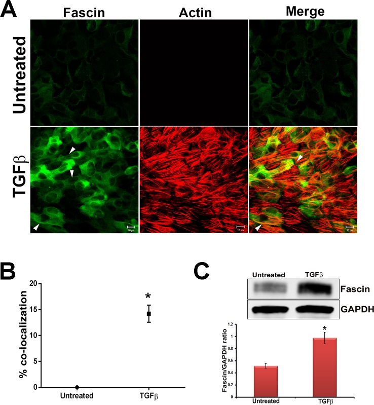
Figure 1.
TGF-β induces fascin expression. (A) Untreated and TGF-β2–treated lens explants were fixed and immunostained for fascin. Fascin-stained slides were costained for F-actin using rhodamine phalloidin. Stained slides were imaged using ×63 water lens of Zeiss LSM510 confocal microscope and analyzed using Zeiss LSM software. Scale bars: 10 μm. (B) A graph for colocalization of fascin with actin was made by scoring at least six different areas of the untreated and TGF-β2–treated lens explants (n = 4, *P < 0.007). (C) Western blot analysis for fascin and GAPDH was carried out using protein lysates extracted from untreated lens explants and TGF-β2–treated lens explants. Densitometric quantification (lower) shows fold increase in fascin normalized to untreated lens explants (n = 4, *P < 0.006).

