Figure 4.
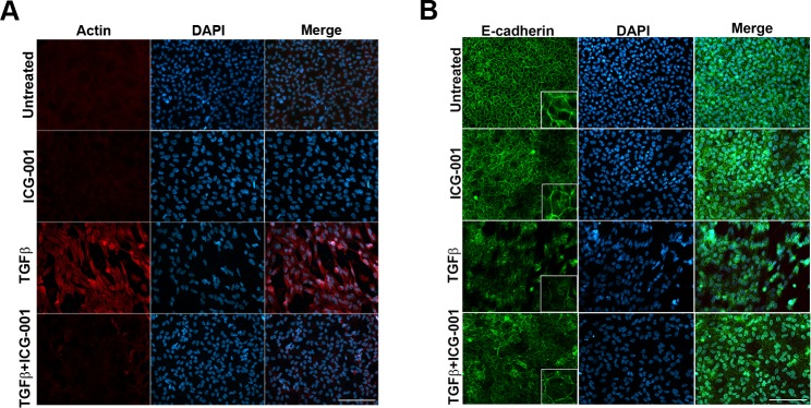
Inhibition of β-catenin/CBP–dependent signaling by ICG-001 prevents stress fiber formation and TGF-β–induced E-cadherin delocalization. LECs incubated with ICG only, TGF-β2 only, and TGF-β2 and ICG-001 immunostained for actin and E-cadherin. Untreated LECs were considered as controls. Stained slides were imaged using ×40 lens of Zeiss Apotome microscope and analyzed using Zeiss Zen software. Scale bar: 100 μm. (A) Actin staining reveals a decrease in TGF-β2–induced stress fiber formation (panel 3) with inhibition of β-catenin/CBP interaction by ICG-001 (panel 4). (B) The TGF-β2–induced loss of membranous E-cadherin (panel 3) was restored by ICG-001 (panel 4).

