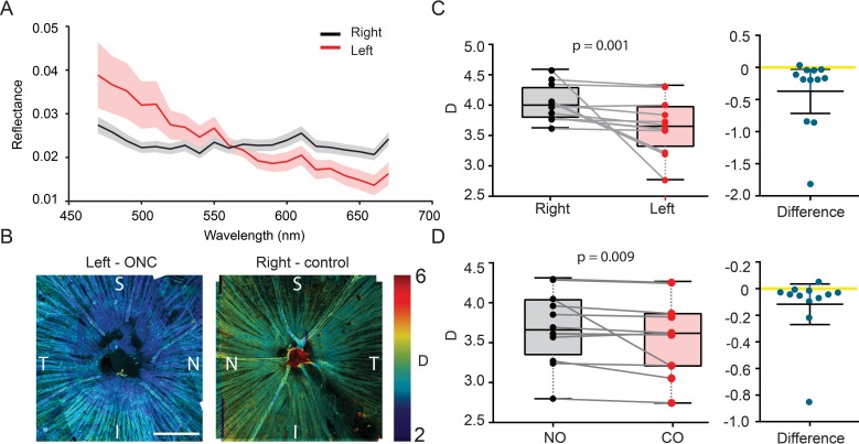
Figure 3.
Ex vivo ultrastructural quantification of early NFL damage. (A) Examples of reflectance spectrum from a left (ONC) and right (control) eye. The shaded areas showed the standard errors. (B) Representative confocal reflectance images of flat-mounted retinas from left (ONC) and right (control) eye from the same animal. D was pseudocolor-encoded on the images. (C–D) Paired comparison of D between left (ONC) and right (control) eyes, and between NO quadrants and CO quadrants; n = 10 mice.

