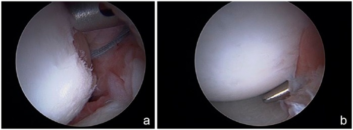
Figure 2.
(a) Arthroscopic view of a right shoulder from the anterior superior portal. A Hill-Sachs lesion is visualized with suture anchor placed through the infraspinatus tendon and capsule and inserted into the posterior aspect of the defect. The drill guide is positioned for the second anterior anchor. (b) After completion of the remplissage, the tendon is approximated at the edge of the articular cartilage defect, effectively excluding the Hill-Sachs defect from the joint.

