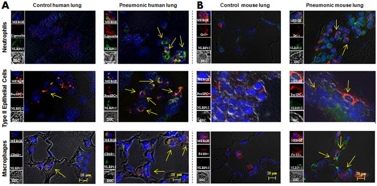
Figure 1. A. NLRP12 expression is increased in myeloid cells (neutrophils and macrophages) and epithelial cells in the lung during ALI/pneumonia.

A. Immunofluorescence microscopy was performed for NLRP12 expression in normal human (control) lung tissue and human lung tissue from bacterial pneumonia. NLRP12 is indicated by green staining, neutrophils are shown by lipocalin staining, epithelial cells are shown by proSPC staining, whereas macrophages are shown by CD68 staining. B NLRP12 expression is enhanced in myeloid cells (neutrophils and macrophages) and epithelial cells in mouse lungs during pneumonia. NLRP12 is indicated by green staining, neutrophils are shown by Gr1 staining, epithelial cells are stained by proSPC staining and macrophages are shown by F4/80 staining. This is a representative image of 5 sections with similar results. Original magnification × 200.
