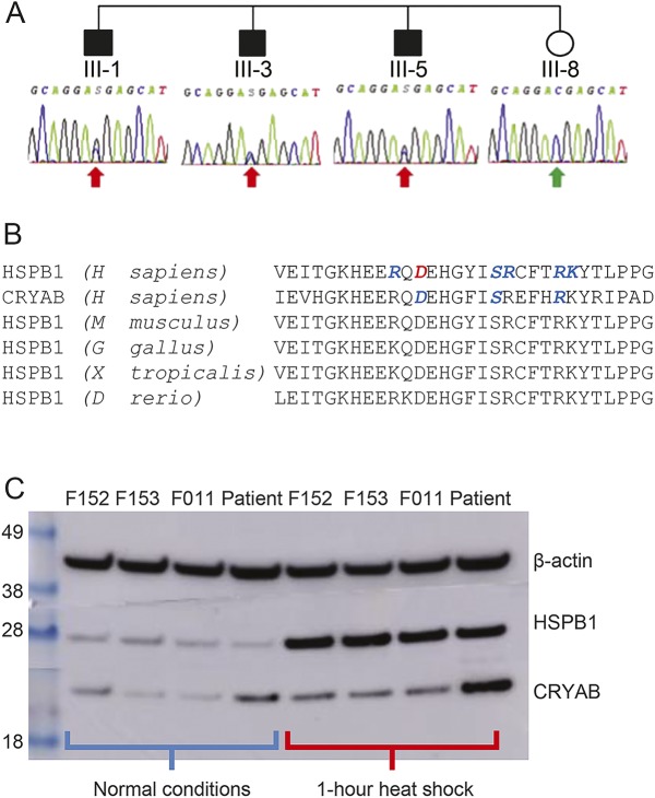
Figure 2. Sequence validation, functional conservation, and Western blot data.
(A) Sanger sequencing electropherograms demonstrating the segregation of the heterozygous HSPB1 c.387C>G variant with the disease phenotype in the 4 siblings. (B) Correspondence of the novel pathologic α-crystallin domain mutation site (in red) in human HSPB1 to human CRYAB and HSPB1 analogs in other species. Sites of known pathologic mutations are in blue. (C) Western blot of HSPB1 and CRYAB expression in control (F152, F153, and F011) and patient-derived fibroblasts before, and immediately after, heat shock by 1-hour incubation at 44°C.

