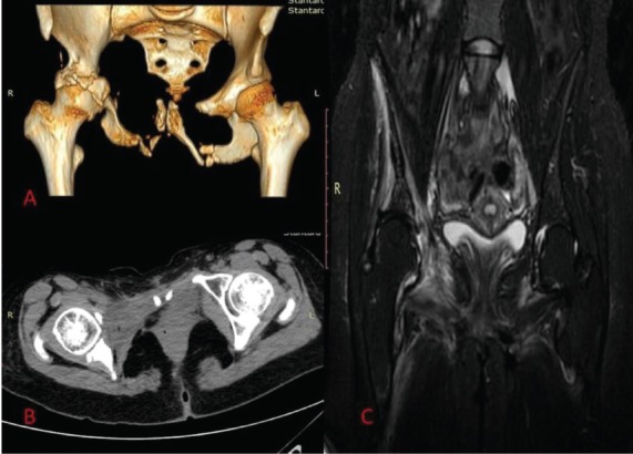
Figure 2.

A) CT scan with 3D reconstruction 7 years after diagnosis. Note the extended osteolysis of the right acetabulum. B) CT scan with T1 weighted images. The right femoral head is completely uncovered. C) Edema of the right hip flexors was revealed in MRI scan.
