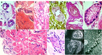
Figure 3. Key morphological changes in patients with vasculitis. EMB: light microscopy (a-i), and electron microscopy (k, l). Skin biopsy (j). Stained with hematoxylin and eosin (a-c, e-j) and Van Gieson (d). Large magnification. Cardiomyocytes with cytoplasmic homogenization, foci of myolysis with formation of cytoplasmic clefts (h), lymphohistiocytic myocardial (i) and endocardial (g) infiltration with deposits of eosinophils, vessels with endothelial proliferation, luminal stenosis, perivascular infiltrates (a-c) and sclerosis (d, e), microvessel ectasia with swollen endothelium and thrombotic lumen occlusion with loose clots (f). Skin: leukoclastic vasculitis (j). Explanation of electron-diffraction photographs - see text.

